
Aiutami a Vincere!
Vota il tuo finalista preferitoFederica Malighetti
Università e Area di Ricerca
Perché votare per me!

Abstract
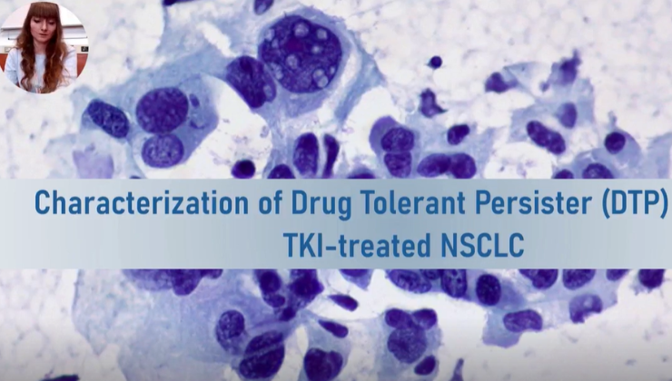
Cancer cell dormancy is a protective state that enables cells to survive treatment. Drug Tolerant Persister (DTP) cells generally lack new genetic mutations but can evade death caused by chemotherapy or targeting agents by entering a slow-cycling phase. Epigenetic modifications, gene expression plasticity, and metabolic flexibility can enable cancer cells to adapt to this challenging environment. In Non-Small Cell Lung Cancer (NSCLC) cells treated with Tirosine Kinase Inhibitors (TKIs), similar mechanisms can lead to the development of drug persistence, allowing the tumor to survive despite treatment. Over time, the adaptation of persister cells may promote the development of drug resistance, making the tumor fully and irreversibly resistant to the TKI. Therefore, the identification and blocking of the mechanisms that support cells during the persistence phase, represent a therapeutic opportunity that could prevent the occurrence of true drug resistance. The aim of this project is to investigate whether drug-tolerant persister cells are present and targetable both in TKI-treated ALK+ NSCLC cell lines and in xenograft models. With the final goal of understanding whether blocking the mechanisms supporting persistence can delay or even prevent the onset of resistance in vitro and in vivo models. The ALK+ H3122 cell line will undergo Alectinib treatment and will be monitored daily by measuring cell confluency.
The construction of RNA libraries from bulk transcriptome will be carried out at baseline (day 0), on day 1 of treatment, on the day when persistence occur and after regrowth, i.e., acquisition of full resistance. The collection of cells that are truly resistant will facilitate a comparative analysis of their transcriptome with that of persister cells. Since persister cells do not undergo clonal selection, whole exome sequencing (WXS) will be performed to ensure that the cells marked as persistent have not accumulated alterations typical of resistant cells. In vitro validations via RT-PCR, western blotting, and genetic manipulations will follow. If the involvement of targetable proteins emerges, we will study the effect of combining Alectinib with drugs that inhibit the identified proteins actively involved in supporting persistence, in both in vitro and in vivo settings. Transcriptomic analysis will provide a comprehensive overview of the pathways that undergo deregulation as tumor cells adapt to Alectinib. It is expected that perister cells significantly modify their transcriptome deregulating various cellular signaling pathways, including those involving drug metabolism, DNA repair mechanisms, and quiescence. In particular, paracrine and autocrine mechanisms are expected to be adopted by persister cells to self-sustain and support neighboring cells. Combining Alectinib with drugs targeting proteins involved in sustaining persistence may exhibit synergistic effects in delaying or preventing resistance. The expected findings may hold significant implications for cancer treatment by unraveling mechanisms of drug adaptation and resistance in ALK+ NSCLC. Understanding these processes can guide the development of more effective therapeutic strategies, potentially extending patient survival. Targeted interventions identified through this research may circumvent and prevent the development of resistance.
Giulia Pedrotti
Università e Area di Ricerca
Università di Verona, area di ricerca neuroscienze, con particolare attenzione alle malattie neurodegenerative rare causate da disturbi mitocondriali.
Perché votare per me!

Abstract
Maternally Inherited Leigh Syndrome is an incurable pediatric mitochondrial disorder with progressive loss of mental and movement abilities, caused by mutations in the ATP6 gene. It encodes for the subunit a of Complex V, which results in its instability, reduced ATP production rate, and elevated ROS levels. Patient-derived neural progenitor cells (NPCs) with a mutation in ATP6 display abnormally high mitochondrial membrane potential (MMP), which has been exploited to identify compounds of the class of PDE5 inhibitors (PDE5i) as possible candidates able to normalize it. However, the action mechanism is still unknown. This project aims to: i) establish a novel preclinical model for MILS to be exploited for a large (> 5500 compounds) high-throughput screening of repurposable drugs; ii) validate the phosphodiesterase 5 inhibitor (PDE5i) Sildenafil as an effective therapeutic option for MILS. Methods: Through a reprogramming-based strategy, human MILS-NPCs carrying homoplasmic mutations on the ATP6 gene were generated.
In my lab, biochemical techniques were used to evaluate CV assembly and enzymatic activity. The MMP of MILS-NPCs carrying different mutations was measured with flow cytometry. Cells were treated overnight with Sildenafil 10 µM, and the pharmacological effect and action mechanism were evaluated. Concomitantly, the MMP of Leigh Syndrome (LS)-NPCs with mutations in complex I and complex IV was investigated with high-content screening (HCS) imaging. Results: MMP hyperpolarization is a constant feature of MILS-NPCs, regardless of the pathogenic mutation, making it an ideal hallmark for phenotypic drug screening. All mutations affected protein stability, impaired assembly of complex V, and reduced OXPHOS-driven ATP content. Sildenafil normalized hyperpolarized mitochondria in MILS-NPCs. LS-NPCs presented depolarized MMP; when treated with Sildenafil, a partial re-polarization was observed. Preliminary data suggest that the action mechanism may involve a cGMP-dependent pathway. Conclusions: Sildenafil normalizes the hyperpolarized MMP and partially rescues depolarized MMP, making it a plausible therapeutic option in LS.
Marta Gino
Università e Area di Ricerca
Università di Torino, Istituto Tumori di Candiolo (IRCCS), ricerca oncologica
Perché votare per me!
Abstract
State of the art The blood vessels in most solid tumors have structural and functional abnormalities (1), which facilitate cancer cell intravasation and dissemination to distant organs. This abnormal vasculature favors resistance to anticancer drugs and radiotherapy, causes chaotic blood flow, and creates local hypoxia that triggers invasive genetic programs (1). Tumor blood vessel defects arise from altered regulation of endothelial cell adhesion to the extracellular matrix (ECM), primarily mediated by integrins. Currently, there is no direct integrin activation tool in live cells able to identify key modulators for therapeutic targeting.
Project’s goals The aim of the project is to unveil novel mechanisms of α5β1 integrin activation in endothelial cells (ECs) by means of: - the generation of a β1 integrin activation conformational sensor (β1IAS) in ECs by CRISPR-Cas9 technology - the identification of β1 integrin activation modulators through siRNA library-based high throughput screening (HTS) and Enrichment Analysis Project The β1 sensor was generated by exploiting the luminescence-based NanoBiT split luciferase tool (by Promega), able to directly and quantitatively detect β1 integrin activation. The two subunits of the NanoBiT probe were cloned into a flexible β1 integrin loop, whose conformation is affected by the hybrid domain swingout that happens when integrin gets activated. Then, a constitutive knock-in (KI) human immortalized Telo Aortic Endothelial Cell (TeloHAEC) clone, stably expressing the luminescent β1IAS, was created by CRISPR-Cas9 technology. The clone expressing β1IAS was used in an endothelial-specific siRNA library HTS, in collaboration with Misvik Biology. The rationale of the screening was to quantify the luminescent signal of the sensor to assess the impact of silenced genes on β1 integrin activation. Genes causing a decrease (z-score<-2) or an increase (z-score>2) in IAS luminescence were identified as, respectively, major positive (111 genes) and negative (44 genes) regulators of β1 integrin activation in ECs. The 155 selected candidates were enriched in pathways related to integrin-based focal adhesion, angiogenesis, and axon guidance. I am currently focused on the functional characterization of the top candidate hits to understand the mechanisms by which they influence integrin activation. This understanding is crucial for leveraging these candidates as therapeutic targets to normalize abnormal cancer vascularization. Recently, a new class of reticular adhesion complexes (RAs), based on clathrin machinery elements, has been reported (2). Since αvβ5 integrin is the primary adhesive receptor involved, future project perspectives aim at the generation of a β5 integrin activation conformational sensor. Methods - NanoBiT is a tool based on a molecularly engineered split luciferase formed by two subunits able to bright luminescence when brought in proximity and after furimazine substrate giving - CRISPR-Cas9 technology has been exploited to insert the NanoBiT gene (555 bp) in human β1 integrin gene - siRNA library-based HTS has been performed on 384-well plates and by lipid-mediated siRNA transfection in β1IAS KI TeloHAECs. Bioinformatic analysis were performed using R language and EnrichR was carried out for enrichment analyses - Immunofluorescence, adhesion/migration assays performed with an electrical impedance-based system (xCELLigence), and protein-protein interaction assays will be employed for candidates functional characterization
I difetti dei vasi sanguigni tumorali derivano da un'alterata regolazione dell'adesione delle cellule endoteliali alla matrice extracellulare (ECM), mediata principalmente dalle integrine. Il mio gruppo di ricerca si concentra sulla comprensione delle basi molecolari dell'adesione delle cellule endoteliali, per modularle e sfruttarle al fine di trovare bersagli terapeutici in grado di normalizzare le dinamiche adesive alterate della vascolarizzazione tumorale.
Nell'ambito del mio percorso formativo, ho frequentato e svolto entrambe le tesi nel gruppo di ricerca di “Cell Adhesion Dynamics” presso l'IRCCS di Candiolo dove ho acquisito competenze tecniche relative alle principali tecniche cellulari, molecolari e di imaging e ho sviluppato un ragionamento scientifico che mi preme continuare a sviluppare. Attualmente sono al secondo anno del Dottorato di Ricerca in Complex System for Quantitative Biomedicine, un programma che mira all'integrazione delle scienze esatte per lo studio e la comprensione di sistemi biologici complessi con un approccio interdisciplinare.
Federica Trenta
Università e Area di Ricerca
Perché votare per me!

Abstract
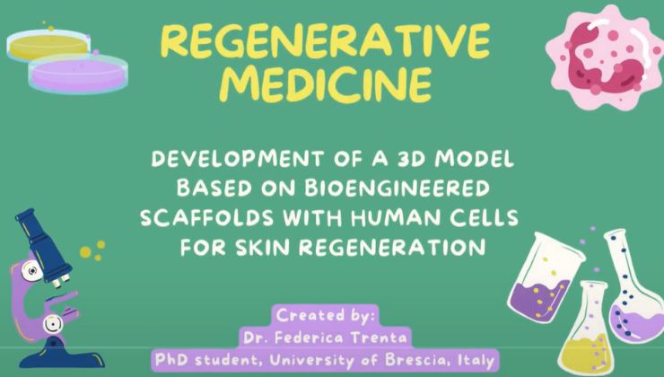
The aim of the project is to develop a 3D model based on biocompatible polymeric scaffolds engineered with human cells for skin regenerative medicine. At present, there is no an equivalent cutaneous able to satisfactorily mimic the native skin, nor a bio-engineered model that is also capable of facilitating the process of skin repair, especially if characterized by a profound loss of substance. In these experimental models, human mesenchymal stromal cells (hMSCs), fibroblast and or keratinocytes can be employed.
The purposes of the PhD project are both the improvement of the scaffolds for cutaneous applications and the optimization and standardization of the cell culture procedures for skin regeneration. Particularly, the 3D polymeric scaffolds will be modified and ameliorated in order to better reply to the clinical needing and these materials will be characterized in terms of porosity, physico-mechanical and thermal properties. Then, the study aims to improve the expansion of hMSCs, from different tissue sources (bone marrow, adipose tissue, umbilical cord), and induce their differentiation for skin regeneration. In fact, the protocols of cells differentiation has to be optimized and standardized, compared to the previously experience, in order to better define the timing for cells differentiation in vitro. In addition, a protocol of fibroblasts and keratinocytes expansion from donor skin biopsy to scaffolds will be settled. Cells will be grown and functionally characterized by comparing the effects of human Platelet Lysate (hPL) versus Fetal Bovine Serum (FBS) addition to culture media into the polymeric scaffolds for skin regeneration. The topical application of hPL can stimulate naturally the tissue regeneration and accelerate the healing processes through the local enrichment of growth factors. Moreover, cell culture procedure using hPL instead of FBS promotes cell differentiation and tissue repair, avoiding the risks of transmitting animal diseases thanks to the crucial mitogenic and chemotactic molecules contained in hPL. Analysis of cells viability, proliferation, differentiation will be done both in the 2d culture, as control, and 3d model in the novel scaffolds.
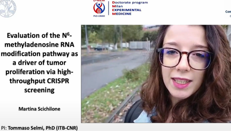
Martina Scichilone
Università e Area di Ricerca
Perché votare per me!
Abstract
The methylation of RNA adenosines into N6-methyladenosine (m6A) is the most abundant internal mRNA modification in mammalian cells. Its deposition and removal are mediated by writer and eraser proteins respectively, while readers target m6A modified RNAs towards different biological processes (e.g., translation and decay). Consequently, m6A affects multiple cellular functions, including cell proliferation. Indeed, CRISPR- knock-out (KO) screenings identified m6A effectors as essential in cancer cell lines, e.g., colorectal cancer (CRC) and acute myeloid leukaemia (AML). This opens new questions regarding the mechanisms by which writers, readers and erasers contribute to the function of m6A in cancer, which may be investigated with CRISPR-cytosine base editing (CBE). In this technology, cytidine deaminase fused to a nicking Cas9 nuclease performs C->T transitions, allowing programmable nucleotide changes. This enables to edit m6A effectors at increased resolution and precision, if compared to classic genetic knockouts.
The aim of this project is to study m6A in cancer cell proliferation by means of a pooled CRISPR screening. By applying CRISPR-CBE and KO, enrichment analysis of the sgRNAs will determine the association between specific single-nucleotide mutations and gene-loss, and the proliferation phenotype. In details, by collaborating with bioinformatic expertise, we prepared the in-silico sgRNA library targeting m6A and control genes. We applied a tiling strategy (i.e., multiple sgRNAs targeting different gene locus) to perform a high-density mutagenesis of target genes. Afterwards, we purchased the synthesis of the library as an oligo pool and its cloning in a lentiviral vector. In parallel, we cloned the CBE and Cas9 sequences into lentiviral vectors that confer doxy-inducibility to their expression. We used these vectors for lentivirus preparation in HEK293TN cells. Following viral isolation, we infected and selected HT-29 CRC lines (HT-29 iBE and HT-29 iCas9). Upon induction, we confirmed the expression at gene (RT-PCR) and at protein level (Fluorescence microscopy and flow cytometry). The next steps include tests on the activity of the editors in selected cells by transfection of a set of sgRNAs targeting test sites. We also planned to prepare the lentivirus from the sgRNA plasmid library and use it to infect HT-29 iBE/iCas9 cells at MOI 0.3 (i.e., one sgRNA per cell). We will select sgRNA+ cells, and, upon induction of the editor (T0), the cells will proliferate for 2 weeks (T1). The Next Generation Sequencing of sgRNAs at T1 versus T0 will result in significantly depleted guides which identify gene domains essential for cell proliferation. Furthermore, we will perform the same experiments in AML cell lines in which m6A pathway proved to be essential for proliferation. Our screening will identify m6A genes important for cancer cell proliferation, and that might represent druggable sites. In addition, we will create a dataset of gene variants associated with the proliferative phenotype, which may be useful for assessing the function of variants of uncertain significance. The study will investigate the role of m6A writers, erasers and readers in CRC and AML proliferation and expect to provide molecular insights for the development of novel drug treatments.
Rita Levi Montalcini
Apri nuovi orizzonti per la tua crescita scientifica!
Il Promega Rising Researchers Award è un concorso nato allo scopo di valorizzare e riconoscere il tuo contributo scientifico e il tuo percorso accademico come dottorando.
Vogliamo supportarti nello sviluppo della tua carriera e aiutarti ad accedere a nuove risorse, conoscenze e connessioni che possano ampliare i tuoi orizzonti.
Per scoprire quali sono i Paesi partecipanti e per avere maggiori informazioni, visita la pagina Rising Researchers Award home page.


Il Rising Researchers Award è importante non tanto per misurare le conoscenze o i risultati, quanto per mettere in evidenza giovani scienziati e le loro idee, che aiutano ciascuno di noi a mettere in discussione i propri limiti. Queste persone eccezionali non sono solo in anticipo sui tempi: ci guidano verso nuove prospettive scientifiche e contagiano tutte le nostre idee.
Poncho Meisenheimer, VP, Research & Development

Il Premio
Il premio consiste in un viaggio presso la sede centrale di Promega a Madison, WI USA, per incontrare il nostro team di Ricerca e Sviluppo e presentare i tuoi progetti.
I voti del pubblico, che decreteranno il vincitore, sono espressi tra il 10 ottobre e il 15 novembre 2024. Il candidato con il maggior numero di voti vince il viaggio a Madison!